Researchers at the University of Oxford have developed a new type of imaging test to provide an early warning of coronary artery disease, and the risk of heart attacks.
The new imaging technique can be applied as a new feature in routine computed tomography angiography (CTA), and will improve the diagnosis and management of coronary artery disease, enabling timely prevention strategies and improving the treatment of thousands of people living with the disease. The findings have been published in the journal Science Translational Medicine.
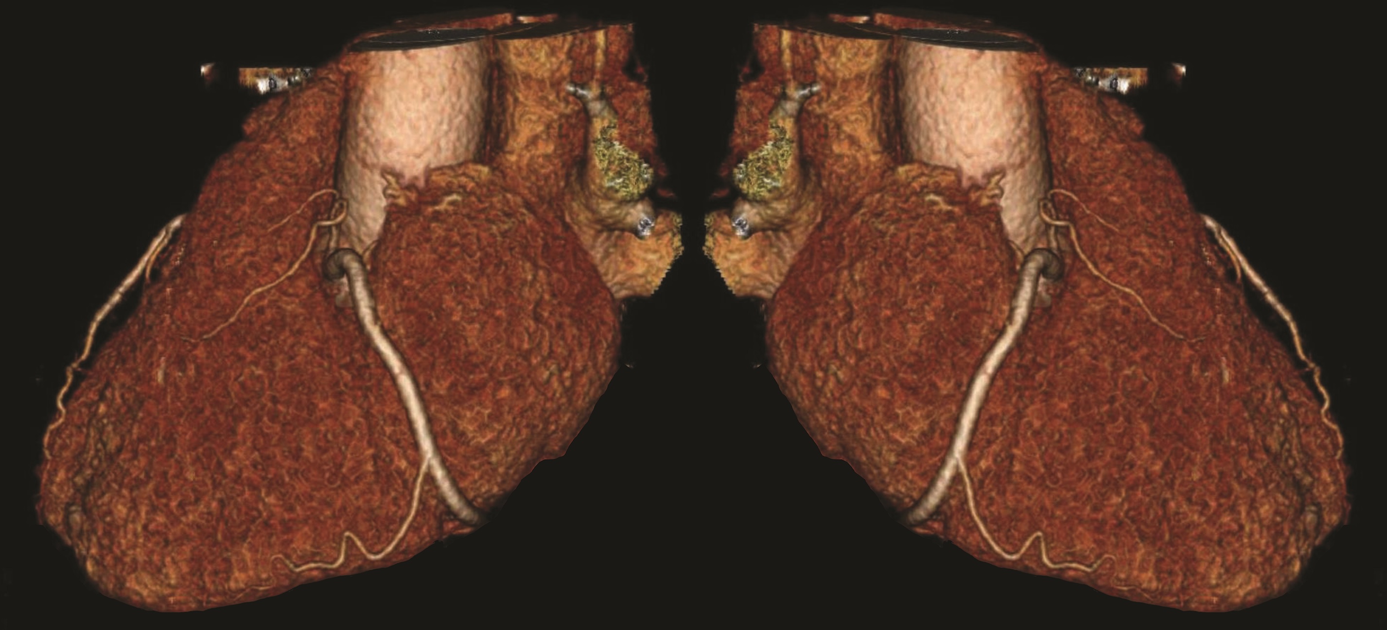
Coronary artery disease occurs when atherosclerotic plaques build up in the arteries that serve the heart. These plaques cause the vessels to narrow, and when they block without warning they lead to a heart attack. Currently, diagnostic methods rely on detecting damage that has already been caused by these plaques – when the damage is irreversible and treatment options are often limited. Inflammation is the local process preceding the development of these plaques, and is also the process that triggers rapid blockages leading to heart attacks. Therefore, detection of inflammation in the heart arteries has been an unmet need in cardiovascular diagnostics for more than 50 years.
The team led by Professor Charalambos Antoniades, Associate Professor of Cardiovascular Medicine at the University of Oxford, discovered a bidirectional communication between the heart arteries and the fat surrounding them. The team found that the fat surrounding these arteries 'senses' inflammation coming from the adjacent artery, resulting in altered fat composition. A new imaging technology, based on routine CTA, called 'perivascular fat attenuation indexing' (FAI), tracks the changes in the fat surrounding inflamed arteries - even in the absence of visible plaques or narrowings.
The technology also detects those 'vulnerable' plaques that are prone to sudden blockages, flagging the individuals at highest risk for heart attacks. Identifying those individuals without narrowings, but with inflamed heart arteries, is expected to allow deployment of targeted preventive measures, early enough to prevent the development of heart disease. Furthermore, detecting patients with established heart disease who have vulnerable plaques prone to lead to a heart attack, will guide application of the new but expensive therapeutic interventions available, to those of the patients who really need them.
Professor Keith Channon, Director of the NIHR Oxford Biomedical Research Centre and one of the co-investigators of the study, said: ‘Developing methods that will enable clinicians to identify people whose vessels are in the early process of developing narrowings, and are at a high risk of causing a heart attack, has been a longstanding goal for research in cardiovascular disease.
‘We’ve known that vascular inflammation drives both the earliest stages of coronary artery disease, and in the events directly preceding a heart attack, but until now we have had no way to easily detect inflammation in the coronary arteries. This new imaging technique can be applied to existing CT angiograms, without additional equipment, making it widely applicable’.
Professor Charalambos Antoniades, who is the lead of the research team at University of Oxford, said: ‘Currently, CT scans can only identify people who have significant narrowings in their heart vessels. But by then the disease may have already caused damage which cannot be undone, and it is not possible to identify which narrowings might progress to cause a heart attack.
‘The new scan offers the potential to find people at an earlier stage of disease and before the damage becomes irreversible. By providing an early warning of disease, the new imaging technique can be used by doctors as the trigger for more powerful treatments designed to reduce the risk of a future heart attack.’
Professor Metin Avkiran, Associate Medical Director at the British Heart Foundation who part-funded the study, said: 'Each year in the UK over 100,000 people die from a heart attack or stroke that has been caused by the rupture of a fatty deposit inside an artery, called a plaque. Discovering which plaques are likely to rupture, so people can be treated before such a devastating event strikes, is a major objective of current research.
'This new method, based on the imaging of cells surrounding arteries, could allow doctors to assess fatty plaques by a non-invasive scan and to see how they change with disease progression or treatment.
'If the technique lives up to its promise in larger trials in patients, it could lead to more effective treatment to avoid a potentially fatal heart attack or stroke.'
Dr James Groves, Technology Transfer Manager at Oxford University Innovation, said: 'Heart disease remains the leading cause of death worldwide, with upwards of eight million victims every year. With the underlying technology behind Caristo Diagnostics, we will have the opportunity to diagnose people at risk of cardiac arrest well in advance of the actual event, giving us an invaluable tool in tackling this global killer.'
The full paper, 'Detecting human coronary inflammation by imaging perivascular fat,' can be read in the journal Science Translational Medicine.